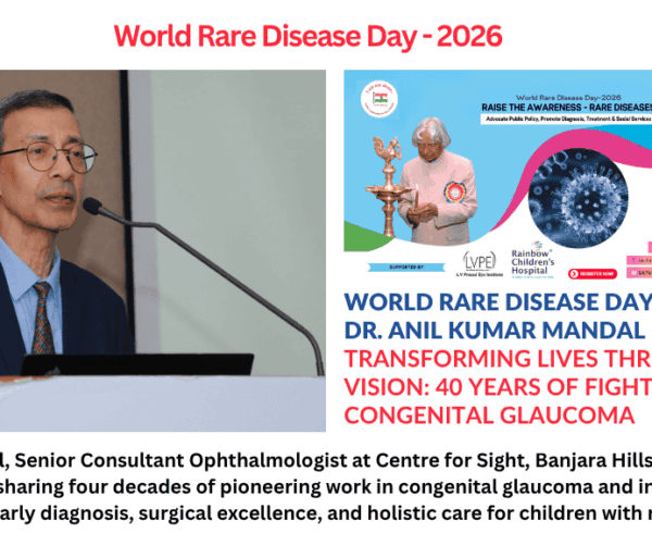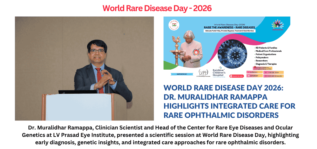
This is a transcribed scientific presentation by Dr. Muralidhar Ramappa, Clinician Scientist and Head of the Center for Rare Eye Diseases and Ocular Genetics at LV Prasad Eye Institute, delivered at the Indian Organisation for Rare Diseases (IORD) World Rare Disease Day 2026 event in Hyderabad, focusing on rare ophthalmic disorders and integrated care. Check the video here.
Hyderabad: We have a Center for Rare Eye Diseases at LV Prasad Eye Institute, which was formally inaugurated about 10 years ago after a soft launch. I have known IORD CEO Prof. Ramaiah Muthyala garu for the last decade, and through this presentation, I would like to pay tribute to him.
Having known him for many years, on behalf of all of us, I would like to thank him not only for inspiring us but also for his unwavering dedication. He does not have to come every year and be present, yet he does so with commitment. It is not a symbolic gesture.
He has been extremely passionate and committed, and every year I have seen renewed enthusiasm to do bigger and better things. I wish him all the success. He has been one of the guiding forces for me in establishing the Center for Rare Eye Diseases at LV Prasad Eye Institute.
It is very important to recognize rare eye diseases. Nearly 600 of them are known, and many cases take an average of nearly 7 years for an accurate diagnosis. Families run from pillar to post without a label for the child’s condition. Without a diagnosis, there is no proper investigation and no treatment. Nearly 95% of these conditions have no definitive cure and are life-limiting.
Some are vision-limiting conditions. Nearly two-thirds are primarily ophthalmic, while one-third are consequences of systemic diseases. We have a battery of investigations to help make a diagnosis.
One key message I want to emphasize is that no single specialist can make a diagnosis in isolation. A pediatrician or an ophthalmologist alone cannot do it. When all specialists—neurologists, geneticists, and genetic counselors—come together under one umbrella, we can diagnose, prognosticate, and provide a roadmap through genetic analysis and counseling. Early diagnosis, early referral, genetic insights, and integrated care can significantly improve the quality of life for many of these children.
Case Study 1
This is a storage disorder. Before corneal transplantation, the child was confined to a room. Even his elder sibling did not like him, as he appeared different physically.
Following transplantation, despite joint deformities, there was a significant improvement. The child, a 9-year-old boy, had multiple systemic issues, including intellectual disability, optic nerve involvement, joint contractures, hepatomegaly, splenomegaly, and a protruding tongue.
Even if survival is limited to 30–40 years, the goal is to improve quality of life.
Case Study 2
At LV Prasad Eye Institute, we encountered a child whose parents noticed abnormal-looking eyes at birth in May 2021. Over time, they searched for answers. Doctors identified a serious corneal condition and referred them from Yemen to India.
At around 3 months of age, the child was examined at LV Prasad Eye Institute, Hyderabad. She was diagnosed with bilateral sclerocornea, with the right eye more severely affected. No surgical intervention was possible for the right eye.
Sclerocornea is a rare condition where the cornea becomes opaque and blends with the white part of the eye, severely limiting vision from birth and making treatment difficult.
The plan was to wait until the child was 4–6 months old for surgery in the left eye. Detailed investigations showed that useful vision might be possible. The team proceeded with a penetrating keratoplasty along with transplantation of limbal stem cells and supportive procedures to improve healing and reduce complications.
Postoperatively, the child was managed with immunomodulators. This specialized approach significantly improves outcomes. Traditionally, keratoplasty in such cases has poor results due to the absence of stem cells and increased risk of graft rejection. However, with this approach, graft survival has improved to 84% over 5 years.
At three years of age, surgery was performed. When the eye patch was removed, the child was able to see for the first time—recognizing her hand and surroundings. This marked a turning point for the family.
Even a failed graft offers better transparency than the original cornea.
Conclusion
In early childhood, the brain is highly adaptable. Meaningful visual experiences during this period help build visual pathways and improve long-term function.
Behind every corneal transplant is a donor and a family who chose to give the gift of sight.
These families often travel long distances, sometimes from war-affected regions, and stay for months to ensure treatment. Ultimately, successful intervention can bring the child to a level comparable with normally sighted peers.
Thank you.